|
VAX-D
and IDD
|
Internal Disc Disruption (IDD)
is a common cause of disabling low back pain in a
substantial number of young, healthy adults. Crock
described this painful entity and reported annular
fissures that distort the internal architecture of the
disc.40 Externally the disc appears relatively intact
and is not deformed. Its etiology has been speculative
but emerging circumstantial evidence favors a traumatic
etiology.
Symptoms
IDD is often found in young adults with a history of
trauma. They present with severe pain that is
predominantly axial in nature (either low back pain or
neck pain), the symptoms are of chronic duration and
typically worse with any loading of the disc and/or
activities that increase the intradiscal pressure. Such
activities often include bending, lifting, sitting and,
sometimes changes in posture. Unlike disc herniations
however, this pain is not the result of disc compression
upon the nerve root.
In most IDD patients the pain does
not usually radiate into either extremity past the knee
to the foot. Typically, these patients have no
"hard/classic" neurological findings. Generally there is
no motor loss, or (dermatomal) sensory loss, no loss of
deep tendon reflexes or root tension signs. |
|
|
Pathology and
Biochemistry of Internal Disc Disruption
IDD is thought to be the result of a structurally
incompetent and internally painful intervertebral disc.
Farfan speculated that IDD was initiated by an endplate
fracture following compression injury to the disc.

He portrayed IDD as the
consequence of an un-arrested repair response that
consumed the disc. The emerging picture is that IDD is
the result of fatigue failure under compression which
results in an endplate fracture that imposes abnormal
stresses on the posterior annulus and which initiates an
internal degradation of the nuclear matrix material. |
Severe axial loading of the spine
may produce end-plate disruption rather than damage to
the annulus fibrosis. This is due to the fact that the
load-bearing capabilities of the annulus are different
than the end-plate and vertebral body. It has been
demonstrated that, as force is rapidly applied, the
end-plate and vertebral body are deformed at a greater
rate than the disc, thus producing disruption in the
end-plate 41.
The region of the vertebral end-plate is innervated by
divisions ofthe gray rami of the sympathetics and
sinuvertebral nerve.42
These nerves travel with blood vessels and have been
noted in all anatomical locations within the vertebral
body except in the deeper zones of the annulus or in the
nucleus pulposus.
Disc material has also been implicated as a causative
agent for chemically induced low back pain due to the
irritating nature of the nucleus pulposus when it comes
in contact with other structures.
43, 44 |
|
Diagnosis of
Internal Disc Disruption
A clinical diagnosis of IDD, in the absence of objective
clinical findings, may be difficult. This is because
there are many spinal problems, which can result in
nearly the exact same symptoms. Because of this,
clinicians must rely on diagnostic testing to arrive at
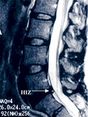
and confirm this diagnosis. Occasionally, a simple MRI
is diagnostic for this process if there is a visible
High Intensity Zone (HIZ), but most of the time it is
non-specific and further testing is needed.
The standard diagnostic test is
provocative discography. This test may show both the
structural abnormality of the disc and may demonstrate
reproduction of concordant pain that is the hallmark of
this disorder. This is performed when surgical treatment
is contemplated. VAX-D is a non-invasive procedure and
the treatment may be undertaken with a diagnosis based
upon history, clinical findings and MRI studies. |
In these patients, the annulus
appears to be extremely painful, although there is no
herniation. They often reveal circumferential and radial
tears radiographically. 45
This is a significant diagnostic factor because the
usual diagnostic tests will appear normal. Not only is
it important to identify the disc level exhibiting IDD,
it is also essential for the discogram to identify
adjacent normal discs if surgical treatment is to be
entertained.
Plain film radiographs are essentially normal, as are CT
examinations and myelography. MRI examination of these
patients may reveal the presence of focal marrow
conversion adjacent to the end-plates of the involved
segments. This may be the result of local stress to the
end-plate region, ischemia, or an inflammatory process.46,
47 |
|
Current
Treatments of Internal Disc Disruption
Since the symptoms of discogenic pain are often related
to increases in intradiscal loading, currently
conservative
 treatment
programs have been aimed at increasing global trunk
strength in an effort to "unload" the disc and thus
decrease the pain, and activity modification. treatment
programs have been aimed at increasing global trunk
strength in an effort to "unload" the disc and thus
decrease the pain, and activity modification.
Many IDD patients do not
achieve satisfactory results with current non-invasive
programs so they often consider surgery. |
The
surgical treatment for internal disc disruption involves
the removal of the painful source or structure, namely
the intervertebral disc. The standard surgical
reconstruction is a lumbar interbody fusion where a
structural bone graft is placed in the evacuated disc
space to allow healing and fusion of the vertebral
bodies.
Often times, a fusion cage, which provides immediate
structural support while serving as a carrier for bone
graft is used to accomplish this. Interbody fusion
obviously does not result in a "normal" intervertebral
motion segment because it fuses the level. |
|
VAX-D
–Treatment For Internal Disc Disruption & Diagnostic
Indicators
Proteoglycans are an essential component of the nucleus
responsible for retention of water content necessary for
metabolic functions of the nucleus. Therefore loss of
proteoglycans accounts for the loss of signal intensity
with darkening of the MRI in T2 weighted images without
concomitant loss in disc height as is seen in the early
stages of degenerative disc disease. This and/or the
presence of a High Intensity Zone (HIZ) on the MRI are
indicative of IDD.
Certain tissue inhibitors of the metalloproteinases
normally keep reparative and destructive activities in
balance. If the balance is disturbed Stromelysin in
particular can outstrip the ability of cells to repair
the annulus and either prevent repair or even create a
necrotic environment where degradation predominates.
While there may be various factors that might contribute
to an imbalance of cellular function it is known that
cell viability depends on a very narrow range of pH
between 6.9 and 7.1. Cellular function in the disc is
dampened by slight decreases in pH to the extent that
only minimal cell function exists if the pH drops below
6.4, a decrease of only 0.6. In this context we have
postulated that a persistent anaerobic state coupled
with accumulation of lactic acid could lead to loss of
cell viability and necrotizing discopathy. Others have
postulated that IDD follows trauma resulting in rupture
of the end plate.
 Cases
of IDD that present with an HIZ, on MRI, are thought to
be a form of necrotizing discopathy with heightened
apoptosis of cellular elements of the disc. This is
especially relevant where the patient manifests
constitutional disturbances consistent with the systemic
circulation of protein degradation catabolites observed
in necrotizing lesions. Cases
of IDD that present with an HIZ, on MRI, are thought to
be a form of necrotizing discopathy with heightened
apoptosis of cellular elements of the disc. This is
especially relevant where the patient manifests
constitutional disturbances consistent with the systemic
circulation of protein degradation catabolites observed
in necrotizing lesions.
In his
textbook on the subject Bogduk commented that necrosis
could be part of the syndrome afflicting some pathologic
processes involving discs.
In considering the mechanisms prevalent in IDD, basic
biochemical investigations are needed to elucidate the
role of |
matrix metalloproteinase (MMP)
inhibitors. There are three principle MMP enzymes
involved in disc degradation, Collagenase, Gelatinase
and Stromelysin. These enzymes are released in an
inactive form and are activated by certain agents such
as plasmin. Collagenase and Gelatinase act together to
cleave Type II collagen primarily found in the nuclear
matrix. Stromelysin is the most destructive, cleaving
various types of collagen found in the nucleus and
annulus as well as aggressively attacking proteoglycans
that constitute the matrix of the nucleus.
Mettaloproteinase degradation, in particular that of
Stromelysin, releases catabolites from collagen and
proteoglycan breakdown which not only can cause low back
pain due to chemical irritation but also can elicit
systemic symptomatology through permeation via a
disrupted annulus into the venous plexus.
Provision of an aerobic medium in the disc necessary for
reparative cell activity could facilitate reversal of
the imbalance of repair vs enzymatic degradation. While
this may be adequate in some cases it can be expected to
fail where enzymatic activity continues uninhibited by
natural biological factors and/or where cell viability
has been damaged beyond recovery.
Many patients diagnosed with IDD will attain temporary
relief of their symptoms with vertebral axial
decompression during a session but they often relapse
after a period of remission. It is thought that they
exhibit an on-going inflammation in the intervertebral
disc associated with degradation. Therefore, for these
patients a definitive VAX-D protocol has been developed
and has proven to be very effective.
Metalloprotienase inhibitors might be directly injected
into the disc. However the ability to create a positive
diffusion gradient through decompression of the disc
might enhance the transfer of orally ingested agents
from the serum into the disc.
Preliminary studies report promise in cases of IDD with
the administration of Methylprednisolone 4-8 mg, given
orally one to two hours before VAX-D. This combined
treatment is recommended each day for the first week of
treatment and continued on Monday, Wednesday and Friday
of the second week. The shortness of this regimen should
not pose any problems with steroid dosage.
Sequential treatment with Doxycycline 200 mg orally may
be given one to two hours before VAX-D. This dosage
should be started on Tuesday and Thursday of the second
week and continued daily for the duration of the VAX-D
therapy.
This compound is utilized here for its activity as a
metalloprotienase inhibitor not as an antibiotic.
Doxycycline was approved as a metalloprotienase
inhibitor by the FDA in 1999. |
|
Summary of VAX-D
Protocol for Internal Disc Disruption
1.
Methylprednisolone - 4 to 8 mg taken orally one to two
hours before each VAX-D session.
 |
First week of VAX-D treatment - One dose each day
|
|
|
Second week of treatment - one dose Monday,
Wednesday and Friday |
2. *Doxycycline
(Vibromycin) - 200 mg. Taken orally one or two hours
before each VAX-D session
|
|
Second week of treatment- Tuesday and Thursday
Thereafter each day for the duration of VAX-D
sessions. |
|
|
For optimum absorption the above medications should
be ingested on an empty stomach, and *Doxycycline
should not be administered to patients who are
allergic to tetracyclines. |
 |
::Continued::
in
Summary
section of Physicians Forum
|


